
Classifying Epithelia
Epithelia: How to classify them
Why do we need to classify epithelia?
The cells in epithelia have different shapes, and different types of epithelia have different numbers of layers of cells (from one to many). The shape of the cells, and their organisation are important to the particular function of each type of epithelia.
How do we classify epithelia?
There are three main criteria for classifying epithelia:
- How many layers of cells are there?
- What is the cell shape?
- Are there any specialisations on the free (apical) surface?
If a specialisation is present, then the type of specialisation present is included in the classification.
Read the classification rules below, then have a go yourself.
How many layers?
One layer - Simple Epithelium

one layer of cells
Find out for more about simple epithelium.
Two or more layers - Stratified epithelium

two or more layers of cells.
Find out more about stratified epithelium
Pseudostratified epithelium

one layer of cells, but the nuclei are at different heights, so it looks as though it is more than one layer. These epithelia usually have goblet cells present.
Find out more about Pseudostratified epithelium.
Cell Shape
When sectioned at right angles to their basement membrane the cells in different epithelia have a variety of shapes:
Squamous:
cells are flat, width is much greater than height.
Cuboidal:

cells appear approximately square.
Columnar:

height is greater than width.
Transitional:
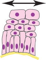
A special form of epithelium, in which the cells can alter their shape. When the epithelium is relaxed they appear cuboidal but when stretched they appear squamous.
Specialisations
Read the summary below, then find out more on the Epithelium Specialisations page.
Microvilli:

Fine, finger-like projections which contain a central core of microfilaments. Increase the apical surface area in cells. Most developed in cells specialised for absorption (e.g. intestinal cells).
Cilia:
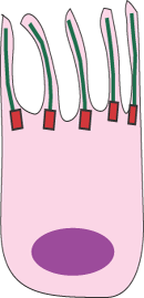
Long, motile projections of the apical surface. These are longer than microvilli. They contain a core of microtubules and beat synchronously. They are found on cells lining the upper respiratory tract for example, where their rhythmic beating moves mucus upwards in the respiratory tract.
Keratin:
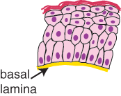
is found in areas susceptible to abrasion and water loss (i.e. skin). Layers of the intermediate filament protein keratin are found on the apical surface.
Goblet cells:
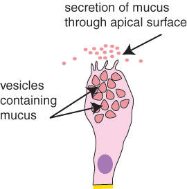
Sometimes epithelia are specialised for secretion - and these cells are called Goblet cells. Goblet cells secrete mucus onto the apical surface. So, they are really 'glandular' epithelia.