
Desmosomes and Hemidesmosomes

Desmosomes are also found in cardiac cells, but the intermediate filament in this case is desmin, not keratin (which is found in epithelial cells). Taken from Molecular Biology of the Cell
Desmosomes connect two cells together.
The picture shows an EM of a desmosome formed between two cells.
Notice the phase dense material between the two cell membranes, which is mad up of transmembrane linker glycoproteins (e.g. demosgleins and desmocollins - which are cadherin proteins).
Also notice the intermediate filaments running from the desmosome into the cytoplasm.
Desmosomes are also known as a spot desmosome or macula adherens (macula = latin for spot), because it is circular or spot like in outline, and not belt- or band shaped like adherens junctions.
Desmosomes are particularly common in epithelia that need to withstand abrasion (see skin - keratinocytes).
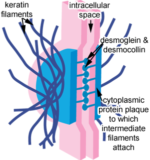 This diagram shows a desmosome. It is made up of a dense cytoplasmic plaque, to which the intermediate filaments attach.
Other proteins run across the membrane into the intracellular space, to connect the two cells together. These 'transmembrane linker' proteins are called desmocollin and desmoglein, which are types of cadherin.
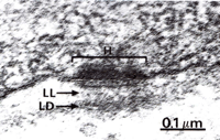
H - hemidesmosome
LL - lamina densa,
LD - lamina lucida - in the extracellular matrix - in this region (the basal lamina) membrane integrins link the cell to the extra-cellular matrix.
Taken from Wheater's Functional Histology.
Hemidesmosomes look similar to desmosomes, but are different functionally, and in their content. connect intermediate filaments to basal lamina.
Hemidesmosomes connect the basal surface of epithelial cells to the underlying basal lamina.
The transmembrane proteins of hemidesmosomes are not cadherins, but another type of protein called integrin.