
Cells: Actin filaments
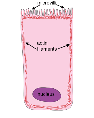 Actin filaments are important in cell shape and cell motility. About half the actin in a cell is unpolymerised. This pool of actin can be released quickly, to polymerise new actin filaments, and push out new projections out of the cell. Actin filaments can polymerise and depolymerise very rapidly in response to cellular signals, changing the cell shape rapidly.
Actin filaments also form a track for myosin motors, which can transport vesicles along the actin track, or interact with actin filaments to contract the cell, as in muscle.
Actin is associated with tight junctions. It's ability to interact with myosin means it can form a contractile network inside the cell, with actin. This is a less organised contractile network than the actin and myosin filament network found in skeletal muscle.
Actin bundles are also found in microvilli.

Actin filaments (microfilaments, about 7nm in diameter) are the smallest filaments of the cell. They are made up of actin monomers which polymerise into filaments, that have two strands which wrap around each other.
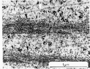
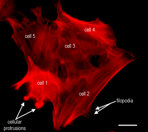
This is a picture of a some cells that have been stained to show the actin filaments. The actin is arranged in filament bundles, and can also be seen in thin projections called filopodia, and in cellular protrusions. The filament bundles would look like those shown on the right, in the EM.
Scale bar, 20 µm.