
Ossification
Bone: Ossification
The formation of bone (ossification) occurs in one of two ways:
- Intramembranous ossification - bone is formed by direct replacement of mesenchyme.
- Endochondral ossification - cartilage model serves as the precursor of bone.
Intramembranous and Endochondral ossification
Both of these forms of ossification are involved in producing most of the bones of the skeleton.
Intramembranous ossification
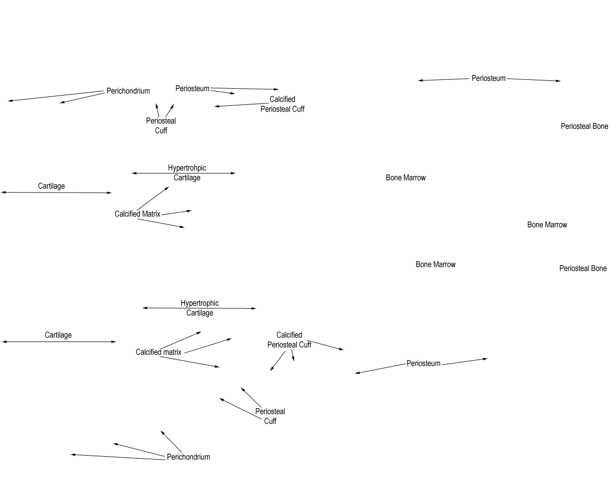
Use the eMicroscope below to examine this longitudinal section through a fetal foot. Find the longer (metatarsal) developing bone. About halfway along, you should see a thin layer of red staining bone matrix close to its surface. This is known as the periosteal cuff.
Make sure you can recognise the periosteal cuff, periosteum, cartilage and perichondrium.
The periosteal cuff is synthesised by connective tissue - i.e. there is no intermediate cartilage stage, but the bone is formed directly onto mesenchyme tissue. This type of ossification is therefore an example of intramembranous ossification.
Endochondral ossification
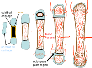
Look again at the section above, in the centre of the diaphysis, beneath the periosteal cuff, the cartilage is being replaced by bone in a so-called primary centre of ossification. At such sites the cartilage begins to undergo hypertrophy and calcification, allowing the penetration of blood vessels which bring with them the osteoblast and bone marrow precursors. This is therefore an example of endochondral ossification.
 In endochondral ossification:
- The bone is formed onto a temporary cartilage model.
- The cartilage model grows (zone of proliferation), then chondrocytes mature (zone of maturation) and hypertropy (zone of hypertrophy), and growing cartilage model starts to calcify.
- As this happens, the chondrocytes are far from blood vessels, and are less able to gain nutrients etc, and the chondrocytes start to die (zone of cartilage degeneration). The fragmented calcified matrix left behind acts as structural framework for bony material.
- Osteoprogenitor cells and blood vessels from periosteum invade this area, proliferate and differentiate into osteoblasts, which start to lay down bone matrix (osteogenic zone).
In the fetus, the primary ossification centre forms first in the diaphysis. Later on a secondary ossification centre forms in the epiphyses.
Cartilage is replaced by bone in the epiphysis and diaphysis, except in the epiphyseal plate region. Here the bone continues to grow, until maturity (around 18 years old). The resulting bone is a thick walled cylinder, that encloses a central bone marrow cavity.
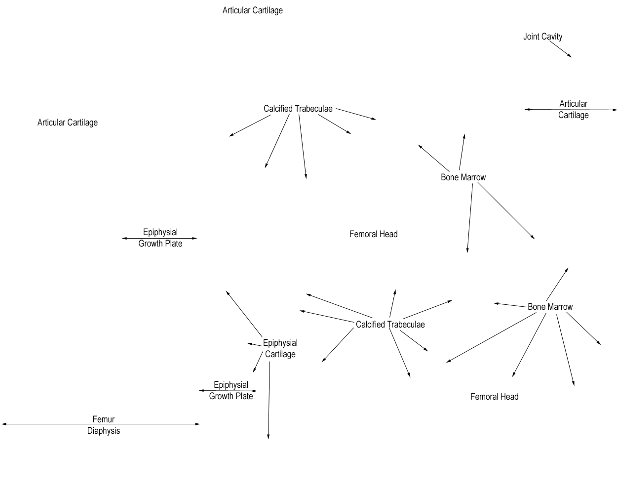
Look at the eMicroscope section below to examine an epiphyseal growth plate.
Make sure you can identify articular cartilage, the epiphysis, the diaphysis, primary and secondary centres of ossification, and the epiphyseal (or growth) plate of cartilage lying between these centres of ossification.To help you recognise the zones in the growth plate, here is a diagram of the different zones, found in the growth plate.
The cartilage is not converted to bone, but is gradually replaced by it. The osteoblasts and blood vessels invade the area of degenerating cartilage.
Find out for yourselves what is happening in each of these zones.
This image may also be viewed with the Zoomify viewer.

Question: What do you think the role of the epiphyseal plate is.
Question: What will happen if it fails in this function?
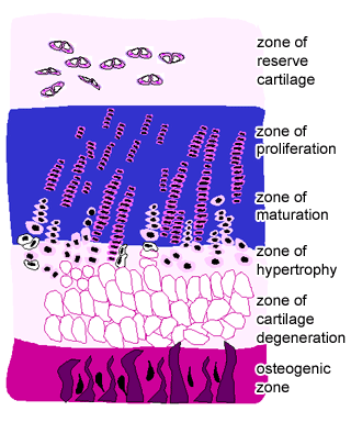 This shows a diagram of the epiphyseal growth plate.