
Epithelium: Pseudostratified
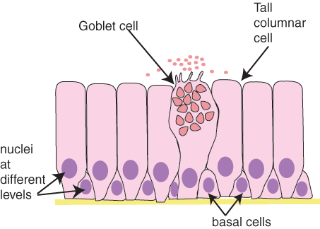 In histological slides of pseudostratified epithelium, it looks as though some of the cells are not in contact with the basal lamina, and the nuclei are at different levels.So, it looks as though there is more than one layer of cells.
However EM shows that all the cells contact the basal lamina, so this is a 'simple' epithelium. But it is called pseudostratified, because of its appearance.
Pseudostratified ciliated columnar epithelium containing goblet cells lines most of the major airways.
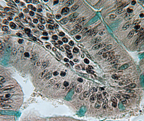 This is an example of pseudostratified epithelium, from the duodenum.
Identify the goblet cells. What shape do the rest of the epithelial cells have? (squamous, cuboidal or columnar?)
It is hard to identify the nuclei of the goblet cells. However, the stain used here makes the goblet cells more easily recognisable, as it stains mucus bright blue (as well as the basal lamina).