
Epithelia: Keratin

Keratin is a type of intermediate filament found in cells.
In the skin, basal cells (next to the basal lamina) divide and move out into the layers above. (For more detail, see the section on skin). As they reach the outer layers, they start to lose their nuclei and cytoplasmic organelles, the cells degenerate and become 'keratinized squames' of the outer keratinized layer.
They finally flake off from the surface (and are a major constituent of household dust).

This is an H&E section of thin skin. There are around 8-10 layers of cells. It's difficult to see the basal lamina in the region of the dividing cells, at the bottom layer.
Notice how the cytoplasm changes colour from the layer of dividing cells at the bottom, to the mature cells in the middle.
Also notice how the cells become more flattened towards the top layer.
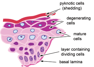
This diagram is taken from the H&E section to the left.
It shows the many layers of cells, and the layer of keratin on the top (outer) surface, the degenerating cells and shedding cells towards the outer edge. The matue cells in the centre are called 'prickle cells'. For more information about this, see the topic on skin.