
Common structure
Basic common structure
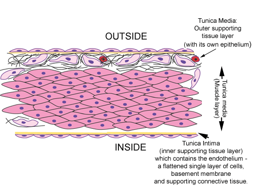 The basic plan of all the blood vessels (except the capillaries) that make up the cardiovascular system consists of these three layers.
- Tunica intima
- Tunica media
- Tunica adventitia
The walls of the cardiovascular system have a single layer of muscle while those of the gastrointestinal tract for example have two, or in some regions three, layers of muscle.
Question: How could you explain this difference?
Tunica adventitia
 This is the outermost coat. It consists of a simple squamous epithelium, basement membrane, connective tissue, blood vessels, and sometimes smooth muscle cells.
This layer needs its own blood supply because it is quite thick. The blood vessels that supply the tunica adventitia are called vasa vasorum (vessels of the vessels).
Tunica media
 This consists of concentric layers of smooth muscle fibres and elastin. Some small blood vessels lack muscle fibres and elastin.
Tunica intima
 This is the innermost coat. In blood vessels the simple squamous lining cells (epithelium) is called the endothelium. The tunica intima consists of the endothelium and underlying basement membrane. A small amount of subendothelial connective tissue and and internal elastic layer (lamina), is sometimes present in some blood vessels.