
- Hyaline - most common, found in the ribs, nose, larynx, trachea. Is a precursor of bone.
- Fibro - is found in invertebral discs, joint capsules, ligaments.
- Elastic - is found in the external ear, epiglottis and larynx.
Types of cartilage
Cartilage: The three types of cartilage
Hyaline cartilage

This is a diagram of hyaline cartilage, showing active chondrocytes sitting in their lacunae
This type of cartilage has a glassy appearance when fresh, hence its name, as hyalos is greek for glassy. It looks slightly basophilic overall in H&E sections.
Hyaline cartilage has widely dispersed fine collagen fibres (type II), which strengthen it. The collagen fibres are hard to see in sections. It has a perichondrium, and it is the weakest of the three types of cartilage.
Look at the eMicroscope of a section of cartilage below. Make sure you can identify chondrocytes, the lacunae, matrix and perichondrium.

This image may also be viewed with the Zoomify viewer.

Fibrocartilage
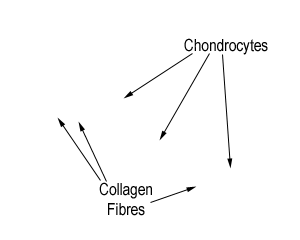
Click image to show/hide labels.
This is a section of an invertebral disc, which contains a layer of fibrocartilage.
Can you find the fibrocartilage in this section? Identify the chondrocytes in lacunae, and thick bundles of collagen fibres.
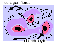 This is the strongest kind of cartilage, because it has alternating layers of hyaline cartilage matrix and thick layers of dense collagen fibres oriented in the direction of functional stresses.
This type of cartilage does not have a perichondrium as it is usually a transitional layer between hyaline cartilage and tendon or ligament.
Elastic cartilage

Click image to show/hide labels.
The picture above is a section of elastic cartilage, stained so that you can see the elastic fibres. In H&E sections, elastic cartilage looks the same as hyaline cartilage, so it has to be specially stained to show the elastic fibres. For example, the Van Giesen stain stains elastic fibres black
In elastic cartilage, the chondrocytes are found in a threadlike network of elastic fibres within the matrix.
Elastic cartilage provides strength, and elasticity, and maintains the shape of certain structure such as the external ear. It has a perichondrium.
This is a diagram of elastic cartilage.

Test your knowledge:
- see if you can identify this cartilage, found in the walls of the trachea.
- see if you can distinguish fibrocartilage from hyaline cartilage