The Heart
The heart is a muscular pump that propels blood at high pressure round the body through the blood vessels. The heart contracts rhymically, and autonomously. Contractions begin at the apex of the heart and spreads through to the postero-basal region.
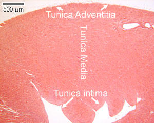
As with the rest of the circulatory system, the heart has three layers, as shown in the diagram below and the photo on the right:
epicardium (tunica adventitia)
myocardium (tunica media)
endocardium (tunica intima)
You also need to know about Purkinje fibres, which lie in the endocardium.
Tunica Adventitia (Epicardium)
This layer contains fibroelastic connective tissue, blood vessels, lymphatics and adipose tissue.
The simple squamous epithelium of the tunica adventitia layer is called the mesothelium
Tunica Media (Myocardium)
In the heart:
The tunica media layer is called the myocardium.
The myocardium is the largest of the three layers, and contains cardiac muscle fibres, and loose endomysial connective tissue that contains lots of capillaries.
Can you identify these two layers in the photograph?
This is picture of the myocardium at a higher magnification.
Now you can see the individual muscle fibres, their striated appearance, and the intercalated discs at the end of each muscle cell.
Tunica Intima (Endocardium)
The endocardium lines the atria and ventricles and covers the heart valves. As well as the endothelium and underlying basement membrane, there is a small layer of loose connective tissue and some adipose tissue.
Can you identify the endothelium, and underlying connective tissue of the tunica intima layer in this photograph?
This diagram shows that the simple squamous epithelium of the tunica adventitia layer of the heart (mesothelium) is also the visceral layer of the serous pericardium.
The pericardium is a two-layered connective tissue sac that encloses the heart. The fibrous pericardium is the outer layer, and the serous pericardium is the inner layer.The space between the two layers is the pericardial cavity, that contains serous fluid. This facilitates the pumping action of the heart.
Background detail about heart contraction:
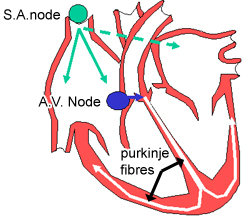
First, impulses are generated by the sinoatrial node (SA), which is found in the wall of the superior vena cava. It is a small mass of specialised cardiac muscle fibres and associated connective tissue, and is supplied by nerve fibres from the autonomic nervous system. Excitation of the SA node sets of a wave of depolarisation around the atria via gap junctions between the muscle fibres.
Next the atrioventricular node (AV) starts impulse generation around the ventricles. The AV node lies in the interatrial septum. Impulses are sent from the AV node into the AV bundle, or bundle of his, which branches to form Purkinje fibres. The AV node is also supplied by nerve fibres from the autonomic nervous system that speed up and slow down the heart rate.
Purkinje fibres lie in the deepest layer of the endocardium and supply the papillary muscles. Hence the apex of the heart contracts first, followed by the papillary muscles, and then the wave of depolarisation spreads up the walls of the ventricles from the base upwards, as shown in the diagram.