Histological Stains other than H&E
| Basic dyes |
| Methyl green |
Green |
| Methylene blue |
Blue |
| Pyronin G |
Red |
| Toluidine blue |
Blue |
| Acidic dyes |
| Acid fuschin |
Red |
| Aniline Blue |
Blue |
| Eosin |
Red |
| Orange G |
Orange |
Acid and Basic dyes
This table gives some examples of basic and acidic dyes used in staining.
For basic dyes, the reaction of the anionic groups of cells (these include the phosphate groups of nucleic acids, sulphate groups of glycosoaminoglycans, and carboxyl groups of proteins) depends on the pH at which they are used.
For acidic dyes, the dye in question can often in addition be selective for particular acidophilic components. I.e. a technique called the Mallory staining technique uses three acidic dyes: aniline blue, acid fuschin and orange G, which selectively stain collagen, cytoplasm and red blood cells respectively.
Periodic acid-Schiff reaction (PAS)
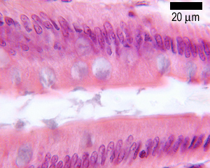
The Schiff reagent is a bleached basic fuschin that reacts with aldehyde groups. This reaction results in a deep red colour in the section. It is the basis of the PAS stain.
PAS stains carbohydrates and carbohydrate rich macromolecules a deep red colour (magenta).
PAS will therefore stain up:
glycogen the intracellular storage form of carbohydrate in cells
Mucus in cells and tissues, Basement membranes, and
Brush borders of kidney tubules and small and large intestines
Reticular fibres (i.e. collagen) in connective tissue and Cartilage.
In the example shown above, The mucin produced by goblet cells is stained a purple colour by this stain.
Masson's trichrome.
This is often used to stain connective tissue. Tri-chrome - means the technique produces three colours. Nuclei and other basophilic (basic-liking) structures are stained blue, cytoplasm, muscle, erythrocytes and keratin are stained bright-red. Collagen is stained green or blue, depending on which variant of the technique is used.
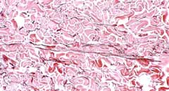
Alcian blue
Alcian blue is a mucin stain that stains certain types of mucin blue. Cartilage is also stained blue. It can be used with H&E, and with van Gieson stains.
van Gieson
This stains collagen red, nuclei blue, and erythrocytes and cytoplasm yellow. It can be combined with an elastin stain that stains elastin blue/black. It is often used for blood vessels and skin.
Reticulin Stain
Stains reticulin fibres blue/black. Used with H&E
Azan
Nuclei are stained bright red, collagen, basement membrane and mucin are stained blue, muscle and red blood cells are stained orange to red. Good for staining connective tissue and epithelium.
Giemsa.
Usually used for staining blood and bone-marrow smears. Nuclei are stained dark-blue to violet, cytoplasm pale blue, erythrocytes pale pink.
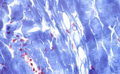
Toluidine blue.
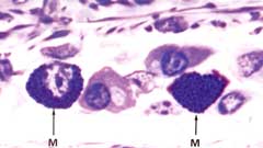
A basic stain that stains acidic components various shades of blue. It is usually used for thin acrylic or epoxy sections.
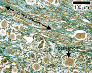
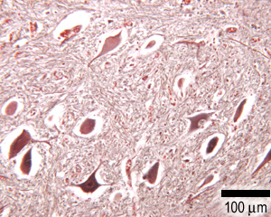
Silver and gold methods.
Sometimes used to demonstrate fine structures such as cell processes in neurones. Produces a black, brown or golden stain.
Chrome alum/haemotoxylin.
Rarely used - stains nuclei blue, and cytoplasm red. For the pancreas, glucagon secreting cells are stained pink and insulin secreting cells are stained blue.
Isamin blue/eosin
Like H&E, but blue is more intense.
Nissl and methylene blue
A basic dye used to stain the rough ER in neurones.
Sudan Black and osmium.
These dyes stain lipid-containing structures such as myelin a brownish-black colour.
Immunohistochemical techniques.
This a specific type of stain, in which primary antibodies are used that specifically label a protein, and then a fluoresently labelled secondary antibody is used to bind to the primary antibody, to show up where the first (primary) antibody has bound. A light microscope, equipped with fluorescence, is used to visualise the staining. The fluorescent antibodies are excited at one wavelength of light, and they then emit light at a different wavelength. Using the right combination of filters, the staining pattern produced by the emitted fluorescent light can be observed. For example, this photo shows some cells that have been immunofluorescently stained for the protein actin.