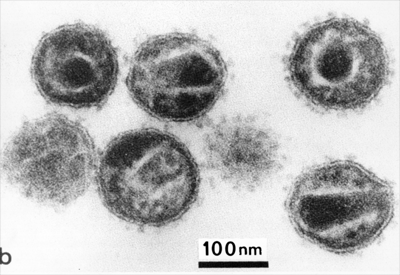
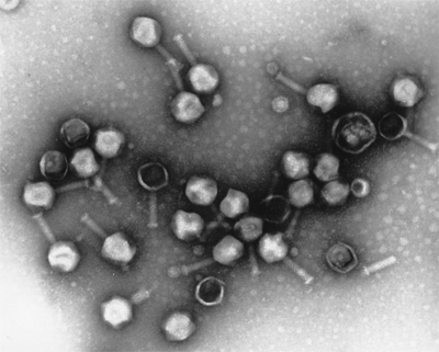
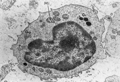
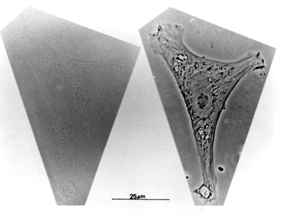
Why do we need to look at cells using an electron microscope?
The limit of resolution of the light microscope is 0.2 µm (greatest magnification is x 1,400).
If we want to see fine detail - objects that are closer than 0.2 µm, then we need a more powerful microscope. This means that we have to use the electron microscope.
Transmission electron microscopes use an electron beam instead of light rays and focuses the light rays with electromagnetic coils instead of lenses. The effective wavelength of a transmission electron microscope, operating at 100kV is about 0.004nm, which is three orders of magnitude shorter than that of light and should, in theory, result in a corresponding increase in resolution. The effective numerical aperture of a transmission electron microscope is 0.012, therefore the resolving power is:
R = 0.004 x 0.61/0.012 nm
R = 0.203 nm.
In practise the resolving power is less than this, mainly because it is impossible to make electromagnetic lenses to the same degree of perfection as glass lenses. Usually with biological specimens, the limit of resolution is around 1-2nm, and the greatest magnification is around 50,000.
Electrons are generated by the cathode (often a heated tungsten filament), and accelerated towards the anode by a large voltage difference, and then focused into a beam with an electromagnetic condenser lens onto the specimen. Electrons that are unscattered by electron dense regions of the specimen reach the objective lens, and the image produced by this lens is further magnified by a projection lens that corresponds to an eyepiece in the light microscope. The final image is projected onto a fluorescent viewing screen, or onto film to produce a transmission electron micrograph.
The tube that the electrons pass through has to be evacuated, for the electrons to travel far enough, and the electron beam is damaging to tissue. For this reason, you cannot look at live cells in the E.M. Even fixed tissue will suffer from electron damage if it is observed for too long.
Find out how we make sections for the electron microscope.


