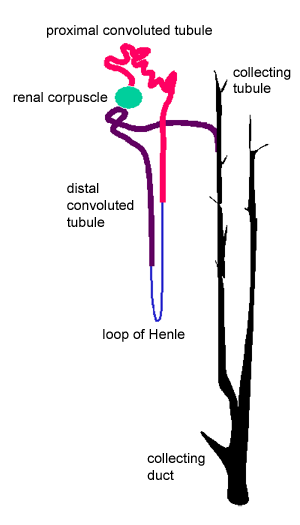
After leaving the renal corpuscle, the filtrate passes through the renal tubule in the following order, as shown in the diagram:
- proximal convoluted tubule (red: found in the renal cortex)
- loop of Henle (blue: mostly in the medulla)
- distal convoluted tubule (purple: found in the renal cortex)
- collecting tubule (black: in the medulla)
- collecting duct black: (in the medulla)

The shape and cross-sectional structure of the different parts of the tubules differs, according to their functions.
First, the proximal convoluted tubule - which is the longest part of the renal tubule - has a simple tall cuboidal
epithelium, with a brush border (microvilli). The epithelium almost fills the lumen, and the microvilli increases the surface area by 30-40 fold.
 What do you think this is for?
Second, the loop of Henle,
This has a thick descending portion (pars recta), a thin descending portion, a thin ascending portion, and a thick ascending portion.
The lumen is made up of simple squamous
epithelium.
This part of the nephron is hard to tell apart from adjacent capillaries, except that there are no red blood cells in the lumen.
Finally, the distal convoluted tubule.
These tubules are less numerous than the proximal convoluted tubules. The epithelial cells are cuboidal, with very few microvilli. The cells stain more palely than those of the proximal convoluted tubule. Click here to see an EM of the DCT.
Collecting tubules are not part of the nephron.
The epithelium of these tubules consist of cuboidal or columnar cells. They empty into collecting ducts that are easy to recognise, because they have large lumens, with pale staining columnar epithelium.
Collecting tubules have two main functions:
1. resorb water in response to the hormone vasopressin.
2. resorb sodium in response to the hormone aldosterone.
From these descriptions, try to find renal corpuscles, proximal
and distal convoluted tubules and loops of Henle
in this eMicroscope section of the cortex from the kidney.
Toggle labels
This image may also be viewed with the Zoomify viewer.


 What do you think this is for?