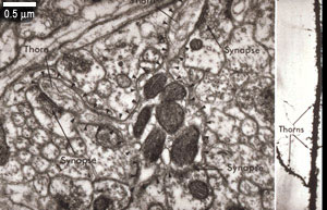
This electron micrograph shows a variety of synapses found in the CNS.
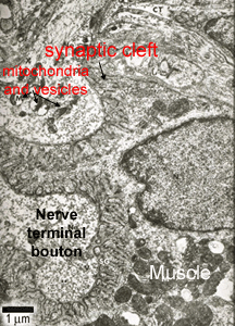
This electron micrograph shows a neuromuscular junction, with two nerve terminal boutons on the left hand side, and a muscle fibre on the right hand side. The folded structure is part of the synaptic cleft formed between the nerve terminal and the muscle fibre. More on the neuromuscular junction.
Synapses are formed between two neurons, or between a neuron and a target cell, such as a muscle cell.
Between two neurons, synapses can form between:
an axon and a dendrite (axodendritic)
an axon and an axon (axoaxonic)
an axon and a cell body (axosomatic)
Chemical synapses are common.
presynaptic terminal - part which delivers the nerve impulse
postsynaptic terminal - part which receives the impulse
synaptic cleft - gap between the pre- and post synaptic membranes.
The presynaptic terminal can be recognised in the EM, because it has synaptic vesicles, that contain neurotransmitter, and mitochondria.
The synapse formed between an axon and a muscle fiber is called a neuromuscular junction. This is a chemical type of synapse.