Nerves: Central Nervous System (CNS)

photograph of spinal cord

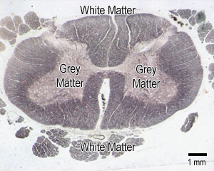
The CNS has a characteristic tissue arrangement called grey matter and white matter.

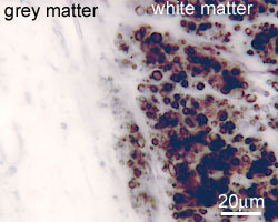
Grey matter contains the cell bodies (perikarya) of neurons and the supporting cells (neuroglia) as well as unmyelinated dendrites.

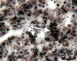
White matter does not contain any cell bodies, but mostly contains myelinated nerve fibres.

The central region of the spinal cord is grey matter, and the surrounding region is white matter.

The picture shows a section of spinal cord, stained by a method that colours myelin blue-black. This is a good way of distinguishing white matter from grey but it can be confusing since it stains white matter black!

After you have finished reading this page, take a look at this section from a diseased spinal cord, and try to work out what's wrong.

  • photograph of white matter

    This is a section of WHITE matter, stained in the same way as the picture above, at a higher magnification. Now you should be able to see all the myelinated nerve fibres.

  • photograph of spinal cord

    This section shows white matter on the right, and grey matter on the left. As the grey matter mainly contains cell bodies, unmyelinated dendrites and supporting cells, it does not stain strongly for myelin, and it looks very pale.

photograph of cell body in CNS

Now look at the picture opposite.

Identify the perikaryon of the neuron.

Can you see that there are some intensely dark purple (basophilic) staining areas in cytoplasm of the cell?

These are known as Nissl bodies or Nissl substance, and are ribosome rich sites of active protein synthesis - accumulations of the rough endoplasmic reticulum.

question
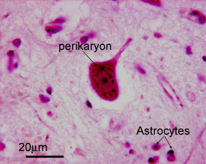
Why do you think there so much Nissl substance present in these cells?

(hint - think about how big the axons are - i.e. how big neurons can get compared to an epithelial cell, for example).

This section also shows the nuclei of some supporting cells, called astrocytes.

You can find out more about supporting cells in the CNS by clicking here.