The peripheral nervous system (PNS) consists of nerves,
which are bundles of nerve fibres, ganglia,
which contain not only nerve fibres but also neuronal cell bodies
(perikarya) and nerve endings.
In the PNS, nerves contain, in addition to fibres, specialised
supporting cells, called Schwann cells. What is their function?
 What are the
Nodes of Ranvier? Why are they important for saltatory conduction?
What are the
Nodes of Ranvier? Why are they important for saltatory conduction?
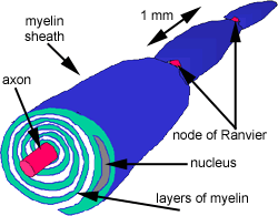
Each Schwann cell wraps its plasma membrane concentrically around
the axon, like a layer of tape, to form a segment of myelinsheath
about 1mm long. (In reality these layers are much more tightly compacted
together than shown here).
The tightly wrapped myelin layers around axons can be seen in the osmium stained section of a peripheral nerve.
Now look at the image opposite. This section shows part of a fascicle
(bundle of nerve fibres) within a peripheral nerve that has
been stained using a technique that stains myelin brown or black. Note that, especially where the fibres have been cut
transversely, myelin sheaths can be seen clearly but that they appear empty because
this technique does not stain the axons that they surround.
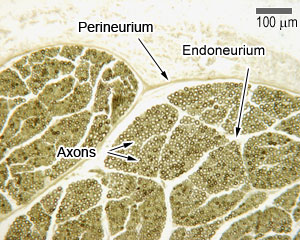
In a typical peripheral nerve such as this there will be several
bundles or fascicles of nerve fibres.
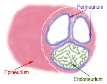
The layers of connective tissue have similar names to those found
in muscle, epineurium, perineurium and endoneurium.
What are ganglia?
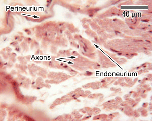
Look at the H & E (pink) stained section that
opposite, and with the help of the labels, see if you can identify
the perineurium and endoneurium.
63x.jpg)