What is a neuron?

diagram of a neuron

Neurons (Nerve cells) are specialised cells that conduct electrical impulses.

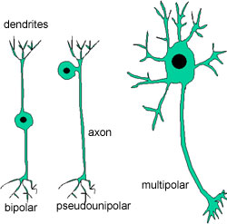
All neurons have the same basic structure:

  • Dendrites extend from the cell body (dendron - greek for tree). These are fairly short, with lots of branches, and they are the points at which nerve impulses are received by the cell.
  • The cell body (perikaryon). Most of the cell bodies of neurons are in the central nervous system (brain and spinal cord), or in the ganglia (which lie just outside the spinal cord) of the peripheral nervous system.
  • The axon: a single nerve 'fibre' which transmits impulses to the distal end. Axons can be very long - around 1 metre, and vary in diameter from 0.2 to 20 µm

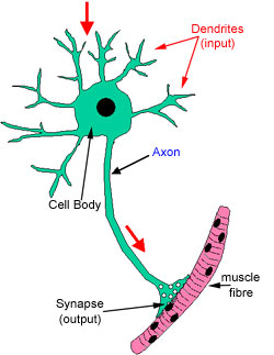
The red arrows show the direction of nerve impulse transmission.

As well as conducting electrical impulses, the cell has to transport proteins, lipids, and other macromolecules, from the cell body to the synapses (anterograde direction), because all of the protein making machinery (ribosomes, endoplasmic reticulum) is in the cell body. This is known as axonal transport. There is a slow transport system of a few millimetres a day, and a fast system, which is about 100 times faster. There is also retrograde flow, in which unused or recycled constituents are returned to the cell body.

(Fast anterograde flow involves microtubules, and the microtubule motor protein called kinesin.)

diagram of neurons

There are three basic shapes to the neurons:

bipolar (single axon and single dendrite) - special neurons in the sensory pathways for sight, smell and balance.

pseudounipolar (single axon and dendrite arise from a common stem) - the primary general sensory neurons are usually pseudounipolar.

multipolar (the commonest) - most motor neurons are multipolar.

photograph of cerebral cortex

This section shows a thick section from the cerebral cortex, stained using Golgi-Cox method, which stains neurons black. Note the variety both of neuronal shapes and of the branching patterns of their processes.

Can you find bipolar, pseudopolar or multipolar neurons?

As most of the nerve cell bodies are either in the brain or spinal cord, or in ganglia, just outside the spinal cord, and the synapses at the end of the axon can be long distances away (i.e innervating muscles at the end of your foot or arm) the axons can be around 1 meter long!

Therefore, because of their large size, and the length and branching of their processes it is difficult to examine the complete structure of an individual neuron in a single conventional sections, as it is unlikely that one section will contain all parts of the neuron.

One way round this problem is to stain the neurons heavily (e.g. by impregnating them with a heavy metal) and to cut a thick section of the tissue which will, with luck, contain all or at least most of some of the cells. This can work for the brain where the neurons can be smaller.