Muscle: Stimulation
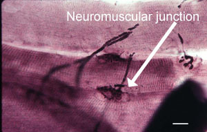
This picture shows nerve fibres approaching skeletal muscle fibres,
and forming a neuromuscular junctions. The structure of the neuromuscular
junction is described in more detail here
Skeletal muscle is stimulated via a nerve impulse, which
depolarises the muscle. However, not all the muscle fibres in the
muscle fibre will necessarily be activated at once. Sometimes, a
subset of muscle fibres is activated, depending on how much force is needed.
When the muscle is stimulated, calcium ions are released from its
store inside the sarcoplasmic reticulum, into the sarcoplasm (muscle ).
Then the calcium ions bind to a protein called troponin, on the
thin filaments, which in turn allows myosin to bind to actin. This
interaction makes the thick and thin filaments slide past each other,
to make the muscle shorten. (see Muscle Sarcomere)
Invaginations of the plasma membrane (sarcolemma)
of the muscle fibres are called T (or transverse) tubules.
The T-tubules lie over the junction between the A- and I-bands (see
diagram).
The two terminal cistemae of the SR together with
their associated T tubule are known as a triad.
Inside the muscle fibre, the T-tubules lie
next to the terminal cisternae of an internal membrane system derived
from the endoplasmic reticulum, called the sarcoplasmic reticulum
(SR), which is a store of calcium ions. Stimulation of the muscle
fibre, causes a wave of depolarisation to pass down the t-tubule,
and the SR to release calcium ions into the sarcoplasm. Calcium
is pumped back up into the SR to lower calcium ion concentration
in the sarcoplasm, to relax the muscle (turn off contraction).
Cardiac Muscle also has T-tubules, and SR. However the T-tubules
lie over the Z-line in cardiac muscle, are less numerous and wider.
The SR is smaller and less elaborate, and stores less calcium ions.
Cardiac muscle cells also depend on extracellular calcium ions,
that enter through the T-tubules and triggers release of calcium
ions from the SR.
Cardiac muscle cells are electrically connected through
gap junctions, so that waves of electrical stimuli pass around
the heart from cell to cell, and all the cells are stimulated to
contract.
Smooth Muscle. The thick and thin filaments are attached
to alpha-actinin in dense bodies (equivalent to Z-lines in skeletal
muscle), which are attached to the plasma membrane by intermediate
filaments. The thin filaments do not have troponin. This
type of muscle responds to a increase in calcium, following nerve
stimulation through a protein called calmodulin. Binding
of calcium to calmodulin, results in the activation of an enzyme
(myosin light chain kinase) that phosphorylates myosin, which activates
it, enabling it to interact with actin.