Epithelium: Stratified Epithelia
Stratified epithelia contain two
or more layers of cells.
The function of this type of epithelium is mostly protective -
the higher the number of layers, the more protective it is. It is
good at withstanding abrasion.
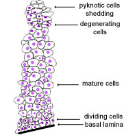
This type of epithelium is constantly renewing itself. Cells in
the bottom layer divide, and the daughter cells move towards surface
maturing and then degenerating.
This type of epithelium can either be keratinising (i.e. the skin)
or non-keratinising (i.e. the oesophagus).
Stratified squamous keratinising epithelium
This type of epithelium is protective against chemical and mechanical
damage, and water loss, and is found in skin, and oral epithelia.
This is an example of thin skin. There are around 8-10 layers of
cells. It's difficult to see the basal lamina in the region of the
dividing cells, in the basal layer. Notice how the cells become
more flattened towards the top (apical) layer. Cells at the top
are flattened and have flattened nuclei - these cells are dying.
Identify the keratin, basal and apical layers.
The oesophagus is an example of a stratified squamous
non-keratinising epithelium.
This epithelium is protective - through its many layers. However,
it does not need the layer of keratin, for protection against drying out.
Many ducts are lined by two layers of layers of columnar/cuboidal
cells, such as the larger ducts of the skin derived glands - sweat glands.
This type of epithelium will provide more protection than a single
layer, but it can't absorb.
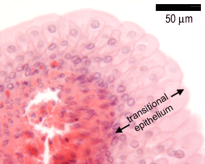
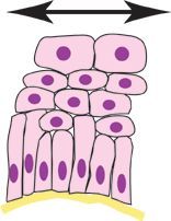
Transitional epithelium is found along almost all of the urinary tract.
Its appearance changes as the epithelium becomes stretched. In
unstretched cells (this is what you would normally see in a histological
section), the rounded superficial cells bulge out (as shown here).