Epithelia: Cilia
Cilia are hair-like motile processes. They are about 10mm
long and about 0.2mm in diameter, longer and
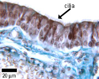
thicker than microvilli. Here you can
see the ciliated cells of the trachea. These cilia are similar in structure
to the cilia found on unicellular free swimming creatures, and on spermatazoa.
The beating of the cilia on cells is used to propel mucus along the trachea,
for example.
Cilia are made of microtubules - shown here in blue. There is a ring of doublet microtubules, and a central pair of singlet microtubules. (See cross-sectional diagram below) At the base of the cilium is the basal body, into which the microtubules are anchored.
This diagram shows a cross-section through a cilium. You can see that arms made of dynein link the doublet microtubules. Dynein is a microtubule motor protein. When the dynein molecules attach to their adjacent doublet microtubule, this makes the cilium bend, via a sliding motion between the microtubules. When dynein releases, the cilium straightens up again. This cyclical action causes the cilia to beat. Dynein uses ATP for energy.