Layers in the Epidermis
This diagram shows schematically, the four different layers found in the epidermis of most skin (thin skin).
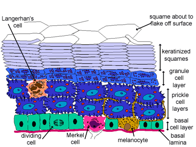
This epidermis of skin is a keratinized, stratified, squamous epithelium.
Cells divide in the basal layer, and move up through the layers above, changing their appearance as they move from one layer to the next. It takes around 2-4 weeks for this to happen. This continuous replacement of cells in the epidermal layer of skin is important. The epidermal layer of the skin (and digestive tract) are the two tissues that are directly exposed to the outside world, and therefore are most vulnerable to its damaging effects. In both, there is constant proliferation of cells in the bottom layer (stratum basale) which constantly move up to the top where they are lost. This means damaged cells are continually shed, and replaced with new cells.
Find out more about the four different types of cell found in the epidermis.
Can you identify the stratum basale, and stratum spinosum in this photo
Stratum basale, and stratum spinosum
The basal cell layer (stratum basale, or stratum germinosum), is a single layer of cells, closest to the dermis. It is usually only in this layer that cells divide. Some of the dividing cells move up to the next layer.
The prickle cell layer (stratum spinosum) is the next layer (8-10 layers of cells). The cells in these layers have lots of desmosomes, which anchor the cells to each other, and contain thick tufts of intermediate filaments (keratin). When the cell shrinks slightly, during fixation, the desmosomes from neighbouring cells remain tightly bound to each other, and these connections look like 'prickles' or 'spines', hence the name prickle cells.
Can you identify the stratum granulosum and stratum lucidum?
Stratum granulosum and stratum lucidum
The granule cell layer (stratum granulosum) is the next layer (3-5 layers of cells). As the cells move up into this layer, they start to lose their nuclei and cytoplasmic organelles, and turn into the keratinised squames of the next layer. The granules contain a lipid rich secretion, which acts as a water sealant.
In thick skin a fifth layer (stratum lucidum) is sometimes identified - between the stratum granulosum and stratum corneum layer. It is a thin transparent layer, difficult to recognise in routine histological sections.
Can you identify the stratum corneum?
This is a photo of the epidermis of thick skin at lower power.
Can you identify the epidermal layers, together with the dermis, and dermal papillae?
Stratum corneum
The keratinised squames layer (stratum corneum) is the final layer. These are layers of dead cells, reduced to flattened scales, or squames, filled with densely packed keratin. In histological sections these cells are flat and hard to see. If soaked in sodium hydroxide they will swell, and then stacks of 10-20 (or even 35) layers of cells packed into hexagonal columns can be seen. The squames on the surface of this layer flake off (making up the main content of household dust).