Four layers of the Gastointestinal Tract
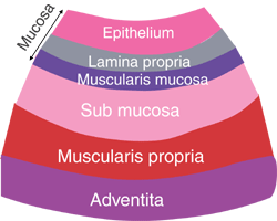
The GI tract contains four layers: the innermost layer is the mucosa, underneath this is the submucosa, followed by the muscularis propria and finally, the outermost layer - the adventitia. The structure of these layers varies, in different regions of the digestive system, depending on their function.
The four layers in more detail:
Mucosa
A lining epithelium, including glandular tissue, an underlying layer of loose connective tissue called the lamina propria, which provides vascular support for the epithelium, and often contains mucosal glands. Products of digestion pass into these capillaries. Lymphoid follicles, and plasma cells are also often found here. Finally, a thin double layer of smooth muscle is often present - the muscularis mucosa for local movement of the mucosa.
Submucosa
A loose connective tissue layer, with larger blood vessels, lymphatics, nerves, and can contain mucous secreting glands.
Muscularis propria (externa): smooth muscle layer.
There are usually two layers; the inner layer is circular, and the outer layer is longitudinal. These layers of smooth muscle are used for peristalsis (rhythmic waves of contraction), to move food down through the gut.
Adventia layer (or serosa)
Outermost layer of loose connective tissue - covered by the visceral peritoneum. Contains blood vessels, lymphatics and nerves.