The production of sperm and eggs/ova (gametes) is a procedure called gametogenesis (spermatogenesis and oogenesis). Gametogenesis involves two rounds of meiotic cell division, in which one diploid cell gives rise to 4 haploid cells.
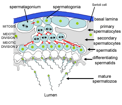
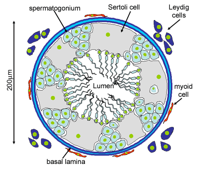
This diagram shows the processes involved in spermatogenesis. The germinal (seminiferous epithelium) of the seminiferous tubules contains spermatogenic cells and Sertoli cells. (Click here to find out about Sertoli cells)
The spermatogenic cells divide by mitosis, then meiosis to form gametes, which mature into sperm by the process of spermiogenesis. Unusually, the developing spermatogenic cells remain connected by cytoplasmic bridges, until they have formed a mature spermatozoan.
There are three types of spermatogonia -
- Type A (dark) which are reserve stem cells
- Type A (pale) which are renewing stem cells
- type B spermatogonia which are differentiating progenitors, and form spermatocytes.
It takes about 2 months for pale A spermatogonia to form new spermatozoa, the total production time for mature human spermatozoa is 74 days.
Compare this process with oogenesis:
In spermatogenesis, each of the four gametes produced by meiosis develops into a mature spermatozoan. What happens to the four gametes in females? (click here to remind yourself).
In spermatogenesis, primitive germ cells are only present in small numbers before puberty. After puberty (driven by testosterone) the spermatogonia multiply continuously to form male gametes. What happens in females?
