Lymphoid: Primary and Secondary Lymphoid Tissues

What is Lymphoid Tissue?
A fluid called lymph, (lymph = clear fluid) flows in lymphatic vessels, lymphatic tissue and red bone marrow.
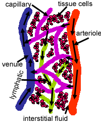
Fluid filters out of capillaries and drains into lymphatic vessels to become lymph.
The content of lymph is the same as interstitial fluid, the fluid around tissue cells.
Lymph eventually drains into venous blood.
Lymph drains interstitial fluid, transports dietary lipids and facilitates immune responses.
What are Primary lymphatic organs?
Primary lymphatic organs are where lymphocytes are formed and mature. They provide an environment for stem cells to divide and mature into B- and T- cells:
There are two primary lymphatic organs: the red bone marrow and the thymus gland. The development of white blood cells (haemopoesis) was covered briefly in the section on blood.
Both T-cell and B-cells are 'born' in the bone marrow.
However, whereas B cells also mature in the bone marrow, T-cells have to migrate to the thymus, which is where they mature in the thymus.
What are Secondary lymphatic organs?
Secondary lymphoid tissues are arranged as a series of filters monitoring the contents of the extracellular fluids, i.e. lymph, tissue fluid and blood. The lymphoid tissue filtering each of these fluids is arranged in different ways. Secondary lymphoid tissues are also where lymphocytes are activated.
These include: lymph nodes, tonsils, spleen, Peyer's patches and mucosa associated lymphoid tissue (MALT).
Filtering lymph
The lymph is filtered by lymph nodes, which are examples of encapsulated lymphoid tissue. There are around 100-200 of these which mostly occur in the neck, thorax, abdomen and pelvis
They contain B- and T-cells, which mostly enter the nodes via the blood stream, and also contains macrophages.
More information about lymph nodes.
Filtering Tissue Fluid
Tissue fluid is filtered by non-encapsulated (or partially encapsulated) aggregations of lymphoid tissue (sometimes called Mucosa Associated Lymphoid Tissue (MALT)). This makes up 85% of lymphoid tissue, in the non-sterile mucosa. They are usually small, around 1mm in diameter, with the exceptions beting the tonsils, peyers patches and the appendix.
These lymphoid aggregations are frequently found close to moist epithelial surfaces e.g mucous membranes of the digestive, respiratory and reproductive systems. Although the epithelia of these tissues has mechanisms to keep bacteria etc out of the body, this is not foolproof. Thus the lymphoid cells in these areas are able to respond to any bacteria or micro-organisms that do get through the epithelia. Activated B-cells in these areas can develop into plasma cells, and produce antibodies, in situ. Lymphocytes from the larger permanent organs (such as the tonsils) are able to patrol the surrounding tissue, and quickly respond to foreign antigens.
Tonsils are large partially-encapsulated masses of lymphoid tissue, found in the walls of the pharynx and nasopharynx, and at the base of the tongue. They form an incomplete ring around the gastrointestinal and respiratory tracts, where they cross over.
Learn more about tonsils.
Peyer's patches are large masses of confluent lymphoid follicles, found in the walls of the ileum, part of the small intestine.
More information about MALT and Peyer's patches.
The appendix is covered in the topic Digestive System, Appendix
Filtering Blood
The blood is filtered by the spleen, another example of encapsulated lymphoid tissue. This is the body's largest lymphatic organ. It is important for antibody production, facilitating immune responses to blood borne antigens, and it also eliminates worn-out blood cells and platelets.
The spleen is a large encapsulated organ in left upper part of abdomen, the outer capsule is fibro-elastic. Click here to find out more.