Small Intestine
Structure
Diagram of a plica circulares.
This is a diagram which shows the villi of the small intestine, as indicated by the arrows in the diagram above, at higher magnification.
The main functions of the small intestine are digestion, absorption of food and production of gastrointestinal hormones. The small intestine is 4-6 metres long in humans.
To aid in digestion and absorption:
- the small intestine secretes enzymes and has mucous producing glands. The pancreas and liver
also deliver their exocrine secretions into the duodenum.
- The mucosa is highly folded.
- large circular folds called plicae circulares (shown in the diagram to the right), most numerous in the upper
part of the small intestine
- smaller folds called villi, which are finger like mucosal projections, about 1mm long.
- the lining columnar epithelial cells have fine projections on their apical surfaces called microvilli.
Together, these folds provide a huge surface area for
absorption. Between the villi there are crypts, called crypts
of Lieberkuhn, which extend down to the muscularis
mucosae. These crypts are short glands.
The lamina propria which underlies the epithelium
has a rich vascular and lymphatic network, which absorbs the digestive
products, and there is a muscularis mucosae layer
immediately at the base of the crypts. The lymphatic capillaries
are called lacteals, and absorb lipids. The vascular capillaries
are fenestrated to aid absorption.
The muscularis externa layer contains two layers
of smooth muscle, an inner circular and outer longitudinal, for
continuous peristaltic activity of the small intestine. There
are around 200 or so lymphoid aggregations called Peyer's patches
in the mucosa.
Look at this photograph of a section through the
small intestine, and make sure you can identify the mucosa,
submucosa, muscularis mucosae, muscularis externa, and villi.
Sometimes the villi are cut in cross-section, and sometimes longitudinally
- and you can see this mixture of sections here.
You can also see part of the fold - plica circulares.
Lymphoid aggregations are commonly found in the sub-mucosa of the
small intestine, an you can see one here. The larger aggregations
of lymphoid tissue are known as Peyer's
Patches.
These patches are more likely to be found in the ileum than in
the duodenum.
Mucosa
On this magnified image of the mucosa of the small
intestine, can you identify: villi, crypts of Lieberkuhn (L), muscularis
mucosa, lamina propria and lymphoid aggregations?
Epithelium and Villi
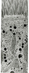
This picture shows a TEM of an enterocyte, showing the villi on the apical surface of the cell.
The epithelium of the villi is made up of tall columnar absorptive
cells called enterocytes, and goblet
cells, which secrete mucin, for lubrication of the intestinal
contents, and protection of the epithelium.
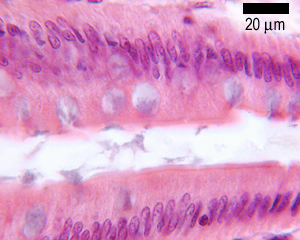
This shows the epithelium of part of a villus at high magnification.
You should be able to identify goblet cells, and enterocytes,
and notice the 'brush border' on the apical surface of the enterocytes,
which is due to the microvilli.
(Click here to compare
the epithelia of the oesophagus, stomach, duodenum, small and
large intestines)
Crypts
The crypts additionally contain
- Paneth cells (at the base of the crypts) - they
have a defensive function, and stain intensely eosinophilic, due
to secretory granules of antimicrobial peptides called defensins,
as well as lysozyme and phospholipase A. These cells last for
several weeks.
- Endocrine cells, (also eosinophilic) which produce
secretin, somatostatin, enteroglucagon and serotonin. One type
of endocrine cell for each type of hormone.
- Stem cells, found at the base of the crypts,
which divide continuously to replace enterocytes (every 2-3 days),
goblet cells, paneth cells and neuroendocrine cells.
Intraepithelial lymphocytes (mostly T-cells).
Duodenum
Look at this picture of a section through the human duodenum.
Identify villi, crypts, muscularis mucosae, mucosa, muscularis externa and Brunner's glands.
The first part of the small intestine is the duodenum,
and its structure is similar to that seen elsewhere in the small
intestine, with some differences. The villi are broader, Peyers
Patches are less common, and it has one unique feature: Brunner's
glands, which are found in the sub-mucosa.
The duodenum is often mistaken for the small intestine, so take
a moment to compare this section to that of the small intestine
in the picture above. Make sure you can distinguish correctly
between the two, and identify Brunner's glands correctly.
Both Brunner's glands, and the goblet cells in the duodenum secrete
mucus. The mucus secreted by Brunner's glands is alkaline,
and helps to neutralise the acid chyme produced by the stomach,
to produce chyme with a pH suitable for the digestive enzymes
of the small intestine.
Digestion:
The chyme is mixed with pancreatic enzymes, and molecules are
absorbed by the enterocytes.
Proteins are denatured and chopped up by pepsin from
gastric glands, and then further broken down by trypsin, chymotrypsin,
elastase and carboxypeptidases in the lumen of the small intestine.
Further enzymes in the plasma membrane of the enterocytes complete
breakdown into amino acids, and each amino acid is actively transported
into the enterocyte.
Carbohydrates are hydrolysed by amylases, and
membrane bound enzymes convert sugars to monosaccharides which
are absorbed by facilitated diffusion.
Lipids are converted into an coarse emulsion
in the stomach, and into a fine emulsion in the duodenum by pancreatic
lipases. Small lipid molecules are absorbed by the enterocytes.