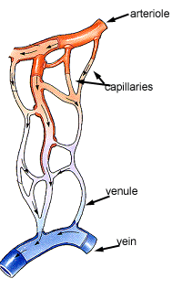
Capillaries
Capillaries are small, normally around 3-4µm, but some capillaries can be 30-40 µm in diameter. The largest capillaries are found in the liver. (capillar comes from the greek for hairlike).
Capillaries connect arterioles to venules. They allow the exchange of nutrients and wastes between the blood and the tissue cells, together with the interstitital fluid. This exchange occurs by passive diffusion and by pinocytosis which means 'cell drinking'. Pinocytosis is used for proteins, and some lipids. Also, importantly, white blood cells can move through intercellular junctions, into the surrounding tissue to repair damage, and fight infections. This route is also used by metastasising cancerous cells.
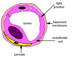
Capillaries have a single layer of flattened endothelial cells, as shown here in the diagram. There are no muscular or adventitial layers. The thinness of the capillaries helps efficient exchange between the lumen of the capillary and the surrounding tissue. Continuous capillaries often have pericytes associated with them. (perivascular cells - peri is greek for 'around') lie just underneath the endothelium of blood capillaries, and are a source of new fibroblasts.
There are three types of capillary:
- continuous
- fenestrated
- discontinuous
Sinusoids, found in the liver can be continuous, fenestrated or discontinuous.
Continuous capillary
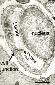
This image is an EM of a continuous type of capillary. Can you identify the two endothelial cells that are bound together by tight junctions. The nucleus of one cell bulges into the lumen of the capillary. The nucleus of the other cell cannot be seen.
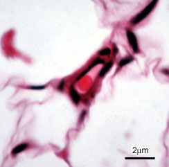
This image is of a capillary in adipose tissue stained using H&E. The delicate capillary wall is supported by fine perivascular connective tissue. Note the single erythrocyte within the capillary's lumen.
Fenestrated capillaries
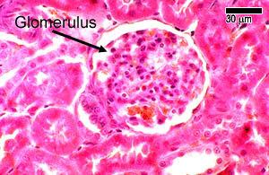
This H&E stained picture shows the convoluted mass of fenestrated capillaries found in a kidney glomerulus.
These are found in some tissues where there is extensive molecular exchange with the blood such as the small intestine, endocrine glands and the kidney. The 'fenestrations' are pores that will allow larger molecules though.
These capillaries are more permeable than continuous capillaries.
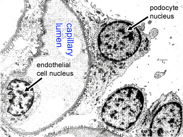
The transmission and scanning electron microscopes below show pores (fenestrae) in the capillary wall of the kidney glomeruli that are not resolved by the light microscope.
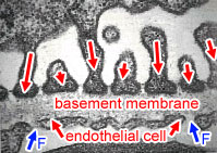
At high magnification, the fenestrations of the endothelial cell can be seen as 'gaps' next the the basement membrane (F) in the picture below.
The red arrows represent the 'podocytes' - foot processes from podocyte (epithelial) cells in the kidney glomerulus.
Discontinuous Capillaries
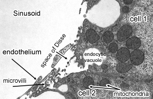
These are only found in the liver. They are formed between the endothelial cells of the sinusoids and hepatocyte cells (Cell 1 and 2 in the picture). The hepatocytes have lots of projections called microvilli that project into the space of Disse. This produces large clefts or spaces between the two layers of cells, that allows proteins, or even blood cells to pass through.
Sinusoids are a special type of capillary that have a wide diameter. These are found in the liver, spleen, lymph nodes, bone marrow and some endocrine glands. They can be continuous, fenestrated, or discontinuous.