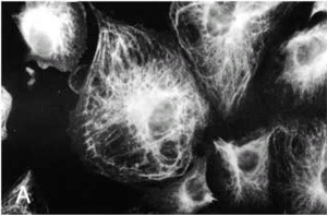
Cytoskeleton

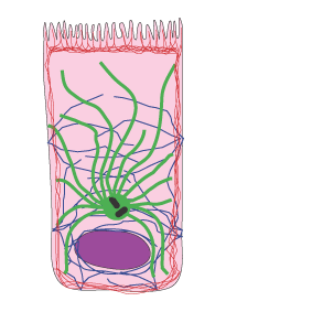
Cells have three types of filaments

  • microfilaments - actin filaments (red in the diagram)
  • intermediate filaments (blue in the diagram)
  • microtubules (green in the diagram).

Collectively, these are known as the cytoskeleton.

diagram of cell with filaments

Actin filaments

Also known as microfilaments, these are the smallest filaments (in diameter) in the cell, at about 7nm in diameter. They are made up of actin monomers which polymerise into filaments, that have two strands which wrap around each other.

drawing of microfilaments

Actin filaments are important in cell shape and cell motility. About half the actin in a cell is unpolymerised. This pool of actin can be released quickly, to polymerise new actin filaments, and push out new projections out of the cell. Actin filaments can polymerise and depolymerise very rapidly in response to cellular signals, changing the cell shape rapidly.

Actin filaments also form a track for myosin motors, which can transport vesicles along the actin track, or interact with actin filaments to contract the cell, as in muscle.

Actin is associated with tight junctions. It's ability to interact with myosin means it can form a contractile network inside the cell, with actin. This is a less organised contractile network than the actin and myosin filament network found in skeletal muscle.

Actin bundles are also found in microvilli.
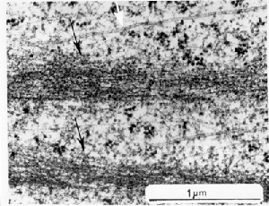
EM of microfilaments

There are two bundles of microfilaments in the picture above (indicated by the black arrows)
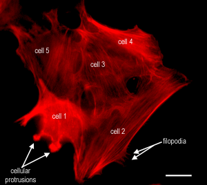
photo of actin in cell

This is a picture of a some cells that have been stained to show the actin filaments. The actin is arranged in filament bundles, and can also be seen in thin projections called filopodia, and in cellular protrusions. The filament bundles would look like those shown above, in the EM. Scale bar, 20 µm.
Microtubules

Microtubules are the largest filaments found in the cell around 20nm in diameter. They are unbranched hollow tubular structures, made of the protein tubulin (see below). The microtubules can polymerise and depolymerise fairly rapidly in cells, as do actin filaments.

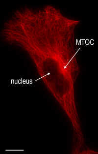
Microtubules are nucleated at (start growing from) the centriole in the centrosome of the cell (The MTOC - microtubule organising centre). The centrosome usually lies just above the nucleus. The microtubules radiate out from the centrosome.

Microtubules are important in cellular transport in interphase cells, as many vesicles are carried along the microtubules by microtobule motor proteins (dynein and kinesins).

Microtubules are also important in mitosis, and are important constitutents of cilia.

diagram of microtubule subunit
Tubulin dimers

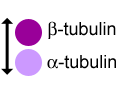
Microtubules are made up of tubulin dimers as shown in the diagram (left). The dimers contain one molecule of alpha-tubulin and one of beta-tubulin. Each dimer is 8nm long.

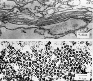
An EM of microtubules

This image shows an EM of microtubules in longitudinal (top) and cross section (bottom), showing their hollow structure.

photo of microtubules in cell

This photo shows the microtubules in a cell. The MTOC (microtubule organising centre) can be seen. (Scale bar, 20 µm). Another fluorescent picture of microtubules, which contains dividing cells.

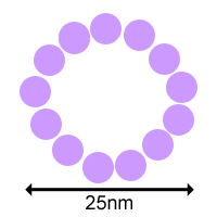
cross_sectional diagram of microtubule
Protofilaments

The tubulin subunits are arranged in rows called protofilaments. These are made up of alternating a- and b-subunits, and the protofilaments assemble into a tubular structure - the microtubule. This tube is 25nm in diameter. The diagram (left) shows a cross section of a microtubule, showing its tubular structure.

diagram of microtubule subunit

This diagram shows how the tubulin dimers assembled into protofilaments, are assembled into the microtubule.

Intermediate Filaments.

These filaments are called intermediate filaments, because they are intermediate in size between actin filaments and microtubules. They are about 8 to 10 nm in diameter.

They act as the cellular scaffold of the cell. They are more stable than either microtubules or actin filaments, and only polymerise and depolymerise relatively slowly in interphase cells. They depolymerise during cell division, and reform at the end of cell division. Also, there are no motor proteins associated with intermediate filaments. (Actin filaments have the motor proteins called myosin, microtubules have kinesins, or dynein). Intermediate filaments are inserted into desmosomes at cell-cell junctions, and help to maintain cell-cell connectivity, as well as the structural integrity of the cell itself. Intermediate filaments can also help to transmit forces between cells, such as in smooth muscle. The type of intermediate filament found in a cell type, is typical of that type of cell. For example:

Epithelial cells contain keratin.

Mesenchyme cells contain vimentin.

Muscle cells contain desmin.

Neurons contain neurofilaments.

Nuclear lamina contain the intermediate filaments called Lamins (A,B and C) in all cells.

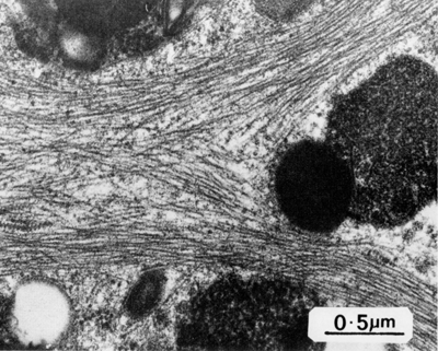
EM of intermediate filaments in a cell

EM of intermediate filaments in a cell

An image of a cell fluorescently stained for vimentin

This is an image of a cell fluorescently stained for vimentin. Taken from Journal of Cell Science 111, 1841-1852 (1998)