Red Blood Cells - Erythrocytes
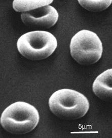
This scanning electron micrograph (courtesy of Dr. Marion J. Barnhart) shows the characteristic biconcave shape of red blood cells.
This is partly due to the arrangement of the actin cytoskeleton that lies just underneath the plasma membrane,
and partly because the nucleus is absent.
This means that the cells can deform easily, and have a high surface area/volume ratio - good for exchange.
Red blood cells are the most numerous type of cell found in blood. One microlitre of blood contains around 5 million cells. They are essential for transport of carbon dioxide and oxygen around the body.
They are 'born' and mature in the bone marrow (see the section on haemopoesis).
When they mature, they make haemoglobin, the protein that binds oxygen. Haemoglobin can also bind carbon dioxide, but at a different site to that for oxygen. Eventually around 90% of the dry weight of the cell is made up of this protein. The nucleus is lost from the cell, phagocytosed by macrophages, and the DNA broken down. The red blood cells can then enter the circulation.
These cells only live for about 120 days. However, the iron in the haemoglobin is extracted from the erythrocytes by the liver and spleen, and the remaining heme is excreted by the liver as bile pigments.
Around 3 million RBCs die and are scavenged by the liver each second.
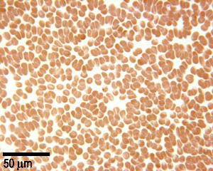
This is a low power image of a blood smear stained with May Grunwald Giemsa stain. This stains the acidic proteins in the erythrocytes pink, as haemoglobin is eosinophilic (acidophilic). All the cells in this image are red blood cells, showing how common they are!
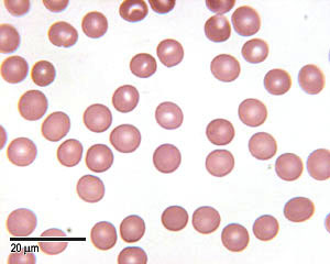
This is an image of red blood cells at higher magnification. The cells are about 7 µm in diameter. At this magnification, it is easier to see the shape of these cells.